

What Three MS Studies Adjusted For

Before a study of multiple sclerosis (MS) can report a difference, it has to settle what the difference is being measured against. A bacterial genus can be counted as a share of the community or as cells per gram of stool. An immune gene's expression can be read on its own or with the patient's weight, smoking and sunlight held constant. A classifier can be scored on the data it was built from or carried to another cell type. Three studies published this year make that second decision visible, because each one changed it deliberately and reported what happened. In all three, something the field had already settled on failed to survive the change, and what remained was smaller.
Counting the Bacteria Instead of Ranking Them
Pauwels and colleagues recruited 228 people with MS in Flanders, 179 of them relapsing-remitting, 69.7% women at a mean age of 46.5 years, and compared them with 2,860 population controls from the Flemish Gut Flora Project recruited in the same region. Alongside sequencing they measured two things most microbiome studies leave out: absolute microbial load by flow cytometry, and stool moisture as a proxy for gut transit time. Both differed. Microbial load was lower in MS, driven by the relapsing-remitting group, and stool moisture was lower, driven by the primary progressive group. In a redundancy analysis across both cohorts, stool moisture explained more of the variation in microbiota composition than the disease itself (2.64% against 0.85%), with age, sex, a general health score, body mass index and faecal calprotectin behind it. Within the MS group alone, moisture explained 3.90%, body mass index 0.88% and serum glial fibrillary acidic protein 0.81%, while MS phenotype stopped being significant.
What Ten Earlier Studies Had Agreed On
Relative profiling returned 45 differentially abundant genera, absolute counts 31, and adding the covariates left 21. The group then set its result beside ten published case-control studies covering 1,065 patients and 874 controls. Akkermansia, raised in MS in several of them and in this group's own relative analysis, was not differentially abundant once absolute counts were used. Roseburia depletion did not persist after correction for moisture, and Faecalibacterium disappeared after confounder control. Three markers held across methods: lower Bacteroides, higher Blautia, higher Methanobrevibacter. Two butyrate producers earlier studies had missed came out depleted, Butyricicoccus and Butyricimonas. Patients carrying the Prevotella community type had lower severity scores, a faster timed walk, and lower triglycerides and glycated haemoglobin. In primary progressive disease, serum glial fibrillary acidic protein correlated inversely with Faecalibacterium and Roseburia. The authors note that the study is cross-sectional and that 16S sequencing cannot reach the metabolic resolution the question needs.
Five Exposures, Each Adjusted for the Others
Falk and colleagues took 121 newly diagnosed, treatment-naive patients in Münster and kept the 96 with complete metadata, 74% women at a median age of 31. Six immune lineages were sorted and sequenced separately, serum proteins and circulating cell subsets were measured in 74 people, and ultraviolet exposure came from satellite records rather than recall. Every association was fitted with all the remaining exposures in the model. Seventy-seven percent of the cohort was vitamin D deficient, 42% were overweight, and body mass index ran inversely to vitamin D. The weight signal sat almost entirely in monocytes, where MVD and SQLE, two enzymes of cholesterol synthesis, were higher and cholesterol homeostasis, oxidative phosphorylation and mTORC1 signalling came up as pathways. In serum, leptin and fatty acid binding protein 4 rose with body mass index while paraoxonase 3 fell, and CD141-positive myeloid dendritic cells rose with it and were also higher in patients with enhancing lesions.
Sunlight and Serum Vitamin D Came Apart
Smoking raised GPR15, a homing receptor, in every lineage except monocytes, and its level in natural killer cells tracked disability. Ultraviolet exposure over the three months before sampling lowered IFI30, LYZ and FCN1 in CD8 T cells and pushed interferon gamma response and oxidative phosphorylation down. Two of them, IFI30 and LYZ, were raised in patients with gadolinium-enhancing lesions at diagnosis, putting the exposure signal and the disease activity signal on the same genes in opposite directions. Serum vitamin D, once the other exposures were held constant, left almost nothing: OCLN higher in monocytes and a correlation with interferon gamma producing natural killer cells. None of the ultraviolet associations in the proteomic data survived multiple testing correction. The authors read the gap as evidence that sunlight's effect in early MS may not run mainly through the vitamin D pathway they measured, and state plainly that supplementation should not be assumed to reproduce it.
Three Genes Out of Twenty-Two Thousand
Xu and colleagues worked from public microarray data rather than a new cohort. The training set held naive CD4+ T cells from 37 untreated people with clinically isolated syndrome and 28 healthy individuals, across 22,017 probe sets. They ran recursive feature elimination with a support vector machine, repeated the selection 100 times, and kept only protein-coding genes picked in more than 85 rounds. Ten genes passed and three were picked in every round: ELOVL1, a fatty acid elongase, GALK2 and PRKCD. A model built on those three and carried to a pan T-cell dataset of 10 patients and 10 controls, then to a mononuclear cell dataset of 12 and 15, reached areas under the receiver operating characteristic curve of 0.84 and 0.83, at sensitivities of 0.90 and 0.91 against specificities of 0.70 and 0.73. ELOVL1 was lower in patients in all three sets. None of the three genes sits in a known MS risk locus.
The Seven Genes That Did Not Travel
Adding the other seven frequently selected genes sent the second test set to 0.49, which is chance, and the first from 0.84 to 0.77; they had been predictive only inside the cell type they came from. Two of them behaved differently. A protein-chemical network linked IP6K2 directly and FUCA1 indirectly to magnesium-ADP, as it had already linked GALK2 and PRKCD, and adding those two lifted the second set to 0.91 and the first to 0.87. The reason for looking there is specific: galactokinase 2, the enzyme GALK2 encodes, phosphorylates N-acetylgalactosamine using magnesium-ATP and releases magnesium-ADP, which opens ATP-sensitive potassium channels. A probe-level check placed the GALK2 difference in the 3' untranslated region rather than the coding exons, so it may be isoform regulation rather than more transcript. The authors list what is missing: small samples, three different cell populations, interferon beta treatment in some test-set patients, and no measurement of magnesium-ADP or channel activity. Across the three studies what changed the answer was not the assay but what the assay was set against.
Disclaimer: This blog post is based on the cited studies and is intended for informational purposes only. It is not intended to provide medical advice. Please consult with a healthcare professional for any health concerns.
References:
Pauwels, A., Devolder, L., Falony, G., et al., D'hooghe, M., & Raes, J. (2026). Deconfounded, quantitative microbiome profiling identifies robust multiple sclerosis markers and clinical covariate associations. Gut Microbes, 18(1), 2681876. https://doi.org/10.1080/19490976.2026.2681876
Falk, S., Schulte-Mecklenbeck, A., Ostkamp, P., et al., Wiendl, H., & Schwab, N. (2026). Distinct multiomic signatures of environmental exposure drive immune dysregulation and disease activity in early multiple sclerosis. Neurology: Neuroimmunology & Neuroinflammation, 13(6), e200636. https://doi.org/10.1212/NXI.0000000000200636
Xu, H., Venäläinen, M. S., Berge, T., & Elo, L. L. (2026). A candidate CD4+ T-cell gene expression panel for multiple sclerosis highlights MgADP-associated genes. Multiple Sclerosis and Related Disorders, 115, 107944. https://doi.org/10.1016/j.msard.2026.107944
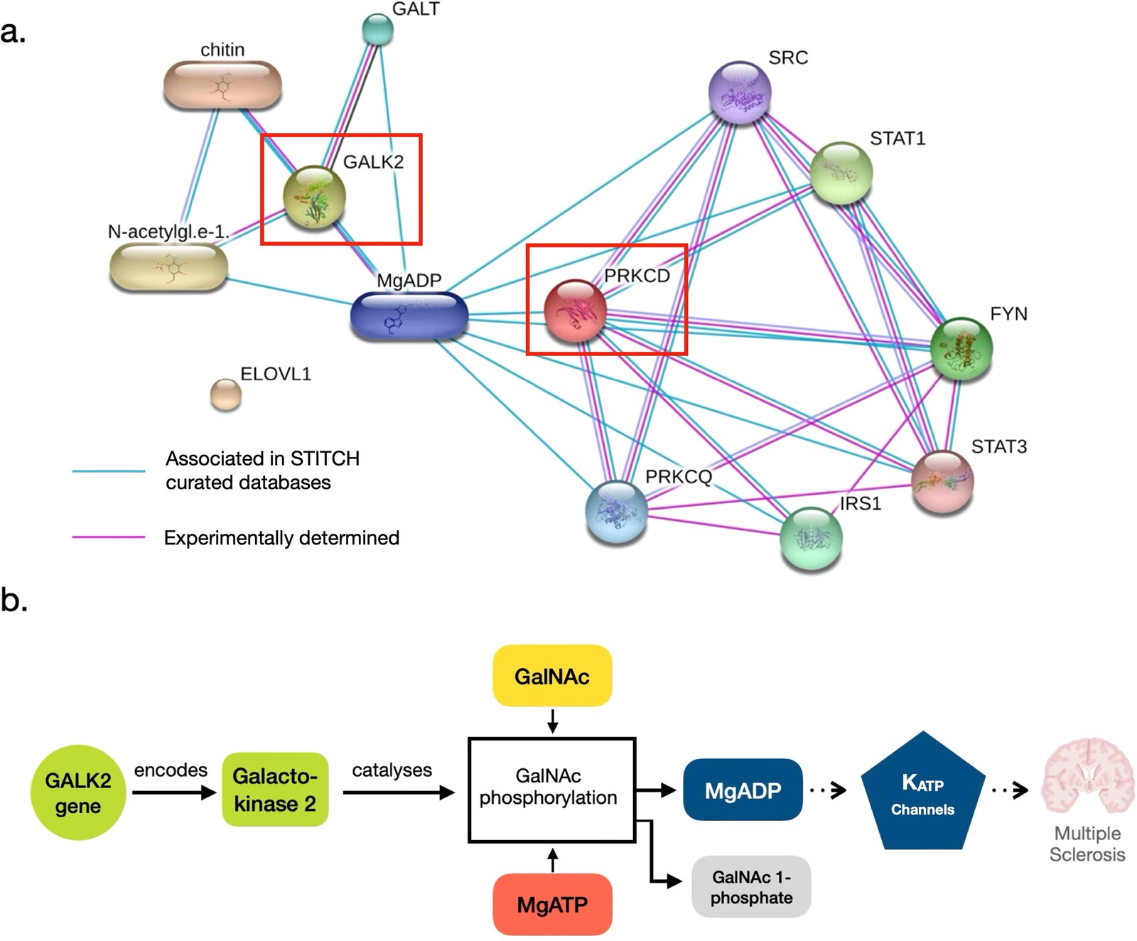
The network that put two of the three selected genes next to magnesium-ADP, and the biochemical chain the authors propose from galactokinase 2 to ATP-sensitive potassium channels. Solid arrows mark established enzymology and dashed arrows the links that were not measured here. (Xu, et. al. (2026), Multiple Sclerosis and Related Disorders 115, 107944, Figure 3, CC BY 4.0)