

When the Control Group in MS Is Not a Healthy Brain
Almost every single-cell study of multiple sclerosis (MS) ends in a gene list, and almost every gene list comes out of one subtraction: patients minus healthy controls. That subtraction is so standard that it is easy to forget it is a choice, and that the genes surviving it are only the genes which differ along that particular axis. Three recent studies change the axis. One compares MS brains against brains from people who had other neurological diseases, then against a fluid rather than a person. One compares women with MS against men with MS. One compares autoimmunity against a spinal injury. The single-cell data is the same kind of data in each; what changes is what it is held up against, and the gene list follows from that.
Ependymal Cells Against Other Diseased Brains
Bigotte and colleagues went after a cell type MS research rarely counts: the ependyma, the ciliated epithelium that separates cerebrospinal fluid from the periventricular parenchyma. They ran single-cell RNA sequencing on fresh human periventricular tissue from four people with MS and four controls, and the controls are the design decision worth reading twice. Three of them had amyotrophic lateral sclerosis and one hereditary spastic paraplegia, so this is MS against other diseased brains rather than against well ones. Against that baseline, 125 cilia-related transcripts were lower in MS ependymal cells and 23 higher, with nine gene ontology terms covering cilium assembly and intraciliary transport coming out reduced. Among the genes going down are CCDC39, CFAP53, DNAI2 and DNAH5, which cause primary ciliary dyskinesia when they carry loss-of-function mutations. In tissue, FoxJ1 protein was lower in MS and fewer ependymal cells carried a cilium at the apex.
Then Against a Fluid Instead of a Person
The second comparison in the same paper swaps the control group for a liquid. Cerebrospinal fluid from people with MS and from people with non-inflammatory neurological disease went onto cultured rodent ependymal cells for 48 hours at a one-to-one dilution, across three small cohorts: four MS against two controls for bulk sequencing, five against two for immunolabelling, seven against three for live recordings of ciliary beating. MS fluid moved 47 cilia-related genes down and 71 up, raised FoxJ1 protein slightly and lowered ciliary beating frequency. Interferon gamma on its own, which is raised in MS fluid, lowered FoxJ1 and slowed the cilia as well. Deleting Ccdc39 from ependymal cells of adult mice shortened and disordered the cilia, enlarged the ventricles at two and four weeks but not at eight, raised periventricular microglial density on the same schedule, and altered nesting behaviour. The authors note themselves that gene and protein changes run in opposite directions across their models and that they cannot resolve the discrepancy.
Women With MS Against Men With MS
LaBarre and colleagues held the disease constant and changed the person. MS affects women roughly three times as often in the United States, while men more often have a severe course at diagnosis, so they asked what separates the two groups inside the same disease. They reanalysed three published single-nucleus datasets of progressive MS brain tissue: 24,890 nuclei from nine donors, 52,323 from twenty-one, and 58,220 from eight. Pooled, that gives 21 MS donors of whom 47.6 percent are women, against 17 controls of whom 35.3 percent are. The subtraction they ran was not MS minus control but MS minus control within each sex, and then those two results against each other, which is a different thing and gives a different answer.
One HLA Gene, Two Effect Sizes
Done that way, the oligodendrocyte lineage moves almost entirely on the female side. HLA-A was higher in MS oligodendrocytes in both sexes, but the magnitude differs by close to fourfold: a log fold change of 3.97 in women against 1.05 in men. Clusterin was higher in female oligodendrocyte progenitor cells and unchanged in male ones. Six electron transport chain genes of mitochondrial origin, MT-ATP6, MT-CO2, MT-CYB, MT-ND2, MT-ND4 and MT-ND4L, were higher in female progenitors and oligodendrocytes and not in the male equivalents. The authors read the clusterin and progenitor results as an environment possibly better primed for repair in women. They are equally direct about what the design cost them: most of the headline findings first appeared in a single female MS sample, so many could be spurious, and only those holding up in the larger dataset were carried forward.
Autoimmunity Against Trauma
Zhu and colleagues changed the comparison in a third direction, holding the molecule constant and changing the kind of injury. They followed the CCL2-CCR2 chemokine axis across spinal cord injury and MS using human cerebrospinal fluid immunoassays, public single-cell datasets, and Ccr2-deficient mice in both a spinal injury model and experimental autoimmune encephalomyelitis. Timing separates the two conditions immediately: a transient surge of CCL2 in acute spinal injury, and no significant movement across MS stages. The single-cell data separates them again. Spinal injury was dominated by rapid innate recruitment that then faded, while the autoimmune model showed sustained accumulation of CD4 T cells and monocytes, with CCR2 acutely induced in infiltrating macrophages after injury and then declining, but persistently high in monocytes and CD4 T cells in autoimmunity. The genotype behaves differently in the two settings too: losing Ccr2 reduced macrophage infiltration after injury with a compensatory expansion of CD4 T cells, whereas in the autoimmune model it suppressed both arms and gave near-complete protection.
What the Comparator Fixes
Lined up, the three show that the comparison group does not sit outside the result; it is part of it. Holding MS ependymal cells against cells from other diseased brains yields a ciliopathy gene set, which a healthy-control comparison would have buried among everything else separating a sick brain from a well one. Holding women with MS against men with MS yields an HLA gene whose effect size depends on sex, a number that vanishes the moment the sexes are pooled. Holding autoimmunity against trauma yields a receptor whose deletion protects in one setting and merely rearranges the infiltrate in the other. None of the three stands in for the usual comparison, and none is offered as a substitute for it. Each answers a different question, and the gene list at the end belongs to that question rather than to MS in general.
Disclaimer: This blog post is based on the cited studies and is intended for informational purposes only. It is not intended to provide medical advice. Please consult with a healthcare professional for any health concerns.
References:
Bigotte, M., Groh, A. M. R., Afanasiev, E., et al., Zandee, S., & Stratton, J. A. (2026). Cerebrospinal fluid-driven ependymal motile cilia defects are implicated in multiple sclerosis. Brain, 149(8), 2654-2667. https://doi.org/10.1093/brain/awaf440
LaBarre, B. A., King, D., Ploumakis, A., et al., Patsopoulos, N., & Chitnis, T. (2025). Sex differences in progressive multiple sclerosis brain gene expression in oligodendrocytes and OPCs. BMC Medical Genomics, 19(1), 13. https://doi.org/10.1186/s12920-025-02294-2
Zhu, X., Liao, Z., Liu, X., et al., Pang, M., & Rong, L. (2026). Context-dependent CCL2-CCR2 signaling drives distinct immune infiltration programs in traumatic and autoimmune CNS disorders. Neurobiology of Disease, 230, 107613. https://doi.org/10.1016/j.nbd.2026.107613
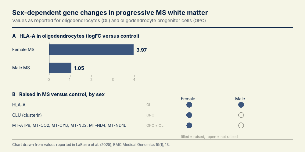
HLA-A rises in MS oligodendrocytes in both sexes but almost four times further in women, while clusterin and six mitochondrial electron transport genes rise only on the female side. Chart drawn from the values reported by LaBarre, et. al. (2025), BMC Medical Genomics 19(1), 13.